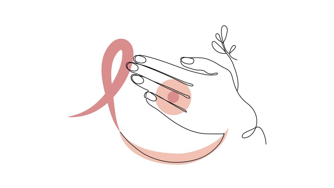
Breast Cancer (Mammary Carcinoma)

Breast cancer, also known as mammary carcinoma, is one of the most common cancer diseases affecting women worldwide. Early diagnosis and targeted treatment are of utmost importance, as chances of recovery significantly improve when cancer is detected in early stages. Breast cancer can cause various symptoms, including changes in the breast or skin, as well as lumps or pain. Since this form of cancer affects many women and is the most common type of cancer in women, regular preventive examinations and personal understanding of the disease are essential.
What is Breast Cancer?
Breast cancer is the most common cancer disease in women worldwide and affects approximately one in eight women during their lifetime. It is a malignant tumor that develops from the cells of the mammary gland or the ductal epithelium. Although men can also develop breast cancer, the risk of disease is significantly higher in women. Initially, there is usually a degeneration of healthy cells in the mammary glands or milk ducts, which can then spread to surrounding tissue and lymph nodes.
Breast cancer cells develop due to genetic mutations that affect cell growth and cell division. These mutations can occur spontaneously or be caused by hereditary factors. One of the most well-known hereditary gene mutations is the BRCA1 and BRCA2 mutation. These genes are responsible for repairing DNA damage. If these genes mutate, the body can no longer effectively repair damage, which can lead to uncontrolled cell growth and the formation of tumors. Women with a BRCA1 or BRCA2 mutation therefore have an increased risk of developing breast cancer.
The incidence of breast cancer increases with age, with most cases occurring between the ages of 50 and 70. Nevertheless, it is not uncommon for younger women to be affected as well. The chances of recovery from breast cancer depend heavily on the stage at which the tumor is discovered. In early stages, when the cancer is still locally limited, the chances of recovery are often very high. When advanced breast cancer is detected, especially if it has spread to the lymph nodes or other organs, the chances of recovery are lower. Nevertheless, adequate treatments in these cases can extend life and improve quality of life.
Types of Breast Cancer
Breast cancer encompasses a variety of different tumor types, each differing in their development, progression, and treatment. The most important distinction is between invasive and non-invasive breast cancer, as this has a decisive influence on the choice of therapy and prognosis. Non-invasive breast cancer, such as Ductal Carcinoma in situ, grows locally and generally has a significantly better prognosis. However, there is an increased risk that a non-invasive tumor may develop into an invasive one. In contrast, the invasive form of breast cancer, such as invasive ductal carcinoma (IDC), penetrates neighboring tissue and therefore requires much more intensive treatment. Depending on the type of breast cancer present, treatment varies from surgical interventions to radiation therapy, chemotherapy, and targeted medications. Survival rates also differ according to the type of tumor. Early, non-invasive forms often have a significantly better prognosis than advanced, invasive carcinomas.
Ductal Carcinoma in situ (DCIS)
Ductal carcinoma in situ, also known as DCIS, is the most common form of non-invasive breast cancer. It represents an early stage of the cancer disease. In this form of breast cancer, the cancer cells are limited to the milk ducts, called ducts, of the breast and have not yet spread to the surrounding tissue. Therefore, DCIS is described as “in situ,” which means “in its original place.” The prognosis for ductal carcinoma in situ is often very good, especially when the disease is diagnosed early. Generally, the chance of recovery is very high when the tumor is detected early. Surgical removal of the affected tissue is the decisive measure for treating ductal carcinoma in situ. Typically, either breast-conserving surgery is performed, where only the tumor is removed, or in some cases, a mastectomy is done, where the entire breast is removed. Depending on the size of the tumor and other risk factors, radiation therapy may be recommended after surgery to ensure that all cancer cells in the affected tissue are removed and to reduce the risk of recurrence. Ductal carcinoma in situ is completely cured in most cases when diagnosed and treated early.
Lobular Carcinoma in Situ (LCIS)
Lobular carcinoma in situ, also known as LCIS, is a less common form of non-invasive breast cancer. LCIS develops when abnormal cells form in the milk-producing glands, known as lobules. However, these cells remain confined to the lobules, which is why LCIS is classified as a non-invasive form of breast cancer. Unlike ductal carcinoma in situ, LCIS is not a direct precursor to invasive breast cancer. However, it does indicate that the affected woman has a significantly increased risk of developing invasive carcinoma—both in the affected breast and in the healthy breast. Although the abnormal cells do not grow invasively, LCIS can be considered a marker for an increased breast cancer risk. The prognosis for LCIS is generally good since it is not an invasive cancer. It is often diagnosed incidentally, as LCIS typically does not cause symptoms and is therefore frequently discovered during routine examinations such as mammograms or biopsies. Due to the increased risk that invasive breast cancer might develop later, regular monitoring of affected women is important. To minimize the risk of subsequent invasive carcinoma, preventive mastectomy may be considered in some cases, especially when the risk assessment is very high. Although such a measure is not necessary for every woman, it may represent an option for women with a particularly high risk to specifically prevent the development of invasive breast cancer.
Invasive Ductal Carcinoma (IDC)
Invasive ductal carcinoma (IDC) is the most common form of invasive breast cancer, accounting for approximately 80% of all breast cancer cases. In this form of breast cancer, cancer cells originate in the milk ducts of the breast and often infiltrate the surrounding breast tissue. This behavior distinguishes ductal carcinoma from non-invasive forms, where the tumor remains confined to the milk ducts.
The signs of invasive ductal carcinoma can vary. Often, a lump in the breast is palpable, which can be detected during self-examination or clinical examination. Skin changes such as redness, retraction, or thickening of the skin over the tumor can also indicate this type of cancer. Some patients also report breast pain or a general feeling of tension in the affected breast.
The prognosis of invasive ductal carcinoma depends on several factors, particularly the stage of the disease at the time of diagnosis and the molecular characteristics of the tumor. These include features such as the presence of hormone receptors on cancer cells that are sensitive to estrogen and progesterone, as well as the presence of HER2, a protein that promotes tumor development. Tumors that have hormone receptors often respond positively to hormone therapy, while HER2-positive tumors can be treated with targeted therapies.
Treatment of invasive ductal carcinoma is therefore typically multimodal, meaning it combines different therapeutic approaches. Depending on the molecular characteristics of the tumor, surgical interventions, chemotherapy, radiation therapy, hormone therapy, and targeted therapies may be considered. The choice of treatment depends particularly on the tumor stage, hormonal receptors, and other biological features of the carcinoma, which significantly influences the prognosis and individual treatment concept.
Invasive Lobular Carcinoma (ILC)
Invasive lobular carcinoma (ILC) is the second most common form of invasive breast cancer, accounting for approximately 10-15% of breast cancer cases. Unlike invasive ductal carcinoma, which originates in the milk ducts, ILC has its origin in the lobules of the breast. The cancer cells spread from the lobules into the surrounding breast tissue, resulting in an invasion of healthy tissue. A characteristic feature of invasive lobular carcinoma is that it often does not form well-palpable, firm nodules. Instead, there is frequently a gradual, diffuse hardening of the tissue. This presents special challenges in diagnosing ILC, as it is often not immediately noticeable and frequently discovered only at an advanced stage. Even in imaging studies such as mammography or ultrasound, ILC may appear less conspicuous, making early detection additionally difficult. The symptoms are therefore often rather non-specific and include changes in breast structure or subtle changes in the skin or breast tissue.
The prognosis of invasive lobular carcinoma is generally worse than that of invasive ductal carcinoma. This can be attributed to the fact that ILC is often diagnosed at more advanced stages, which can limit treatment options and the likelihood of cure. ILC often grows more slowly than IDC, meaning it is less aggressive in many cases. The prognosis is highly dependent on the size of the tumor, the stage at the time of diagnosis, and the biological characteristics of the tumor, such as hormone receptors or the presence of HER2.
The treatment of invasive lobular carcinoma is similar to that of invasive ductal carcinoma and typically includes surgical removal of the tumor, followed by supplementary treatments such as chemotherapy, hormone therapy, and possibly radiation therapy.
Triple Negative Breast Cancer
Triple negative breast cancer is a particularly aggressive form of breast cancer that accounts for approximately 10-15% of all breast cancer cases. This form occurs more frequently in younger women and is characterized by the absence of three specific receptors that play a central role in the treatment of other breast cancer types. “Triple negative” specifically means that the tumor does not express estrogen, progesterone, or HER2 receptors on the surface of cancer cells. This has profound implications for the treatment and prognosis of the disease. Since these receptors are not expressed, conventional targeted therapies such as hormone treatments that target estrogen or progesterone receptors, and HER2 therapies used for HER2-positive tumors, are not effective for triple negative breast cancer. Therefore, treatment relies primarily on chemotherapy. The goal of chemotherapeutic drugs is to destroy cancer cells through cell division and growth. However, this type of breast cancer does not respond equally well to chemotherapy in all cases, which complicates treatment and makes the prognosis challenging. Triple negative breast cancer is generally more aggressive than other forms of breast cancer. The tumor often shows a tendency for faster growth and spread to adjacent tissue. The recurrence rate for triple negative breast cancer is particularly high, meaning that after treatment, there is a greater probability that the cancer will return. Additionally, survival time for women with triple negative breast cancer is often lower, especially if the tumor is discovered at advanced stages.
Therefore, the prognosis for triple negative breast cancer is often worse than for other, receptor-positive forms of breast cancer. However, it is crucial to emphasize that not all patients with triple negative breast cancer follow the same course. Features such as tumor size, metastasis formation, and the general health situation also influence the prognosis.
Inflammatory Breast Cancer
Inflammatory breast cancer is a rare but extremely aggressive form of breast cancer that accounts for only about 1-5% of all breast cancer cases. This type of breast cancer differs significantly from more common forms as it affects the lymphatic vessels in the breast and causes severe inflammatory symptoms. The most typical symptoms are intense redness, swelling, and warmth of the affected breast, as well as thickened skin with dimple-like depressions referred to as “orange peel skin.” These skin changes, caused by the blockage of lymphatic vessels by cancer cells, are a characteristic feature. Since inflammatory breast cancer is often confused with an acute inflammation, diagnosis frequently occurs only when the tumor has already reached advanced stages. As a result, the prognosis compared to other types of breast cancer is often worse, as the cancer has often already spread locally or metastasized to other regions of the body by the time it is discovered. The growth of tumors in the lymphatic vessels contributes to the rapid spread of the disease and therefore makes early diagnosis more difficult. Inflammatory breast cancer is typically treated with a combination of chemotherapy, surgical intervention, and radiation. To reduce the tumor size and slow the spread of cancer cells, chemotherapy is often used before surgery. Since the tumor often shows widespread distribution, in many cases a mastectomy (removal of the breast) is combined with supplementary radiation therapy. To increase patients’ chances of survival, intensive and immediate therapy is necessary for this aggressive form of cancer. The prognosis for patients with inflammatory breast cancer remains more challenging than for other types of breast cancer despite advances in treatment. Early diagnosis and individually tailored therapeutic approaches are crucial to control the disease and reduce the risk of further spread.
Causes and Risk Factors of Breast Cancer
Breast cancer is the most common type of cancer among women. While the exact causes of breast cancer are not yet fully understood, there are several known risk factors that can increase the likelihood of developing the disease. These include genetic, hormonal, and lifestyle factors:
- Female gender: Breast cancer occurs significantly more often in women than in men.
- Age: The risk increases with advancing age, particularly after age 50 and after menopause.
- Family history and genetic factors: Mutations in the BRCA1 or BRCA2 genes significantly increase the risk. Other genetic alterations may also play a role.
- Early menarche and late menopause: An early onset of the first menstrual period, before age 12, or a late menopause, after age 55, increases the risk of breast cancer, as the breast tissue is exposed to hormonal influences for a longer period.
- Postmenopausal hormone therapy: Hormone therapy for more than five years can increase the risk, especially when estrogens and progestogens are taken in combination.
- Alcohol consumption and smoking: Both can increase the risk of breast cancer through oxidative cell damage and hormonal changes.
- Overweight and obesity: A high percentage of body fat is particularly problematic after menopause, as fat tissue produces estrogens that can increase breast cancer risk.
Many of these risk factors can be actively influenced, particularly through a healthy lifestyle. A balanced diet, regular exercise, and avoiding smoking and excessive alcohol consumption can help reduce the risk of breast cancer. Especially after menopause, it is important to avoid being overweight, as fat tissue produces estrogens that can promote the growth of cancer cells. In addition to a healthy lifestyle, early detection also plays a crucial role. Regular preventive examinations, such as mammography screening, can help detect breast cancer at an early stage, when the chances of cure are highest.
Breast Cancer Symptoms and Signs
Breast cancer can manifest through various symptoms and signs that cannot always be clearly identified. Some changes in the breast are often immediately visible, while others frequently remain unnoticed or are misinterpreted. Breast cancer often causes no pain in early stages, which consequently leads to its late detection. However, there are certain warning signs that can indicate a potential illness. These include lumps or hardening in the breast area, skin changes, an inverted nipple, as well as less known symptoms such as chronic fatigue or non-specific complaints. It is particularly important to distinguish these symptoms from non-specific cancer symptoms like general discomfort, mild pressure sensation, or occasional pain, as these can also have other causes.
Lumps or Hardening of the Breast
The most common sign of breast cancer is a palpable lump or hardening of the breast. Many affected individuals notice the change by chance or usually during a self-examination. Typically, lumps that indicate a malignant change are irregularly shaped, relatively firm, and difficult to move. In contrast, benign cysts or fibroadenomas are often clearly definable and easier to move. In most cases, these lumps are painless, which is why they are not initially viewed as threatening. In addition to hardening in the breast tissue, swollen lymph nodes in the armpit can also indicate tumor spread. Such lymph nodes often appear enlarged, firm, and immobile, which may suggest possible involvement. Regular breast self-examination is an important method for early detection. Both breasts should be systematically palpated. The goal is to notice new, unusual lumps or tissue changes early. However, this method does not replace medical examination or imaging procedures such as mammography, which can make the smallest changes visible before they are palpable. If you notice an abnormality, we advise you to consult a doctor as soon as possible for a more detailed assessment.
Swelling of Regional Lymph Nodes
Lymph nodes play a central role in the immune system, as they filter pathogens and abnormal cells from the body. In breast cancer, swelling of regional lymph nodes can occur when cancer cells spread through the lymphatic system. Particularly affected are the axillary (in the armpit), supraclavicular (above the collarbone), and infraclavicular (below the collarbone) lymph nodes. Affected lymph nodes are characterized by painlessness, firmness, and limited mobility. Inflamed lymph nodes, on the other hand, are often tender and mobile, while those invaded by cancer cells typically have a harder consistency. When significant swelling is observed, this may indicate that the tumor has already spread beyond the breast, with metastases present in the lymph nodes. Additional self-examination of the armpit can help detect suspicious changes early. However, swollen lymph nodes are not always a sign of breast cancer; they can also be caused by infections or other benign processes.
Changes in the Nipple
Changes in the nipple can be an important indication of breast cancer, especially when they occur suddenly and cannot be explained by hormonal fluctuations or mechanical stimuli. An inverted nipple, unusual discharge, or hardening in the nipple area should always be medically evaluated. An alarming symptom is the sudden inversion of the nipple. While some people naturally have inverted nipples, a new, unilateral retraction may indicate a tumor. This occurs because cancer cells invade the surrounding tissue and reduce the milk ducts or supporting tissue of the nipple, pulling it inward. Another significant symptom is bloody or clear discharge from the nipple that is not associated with pregnancy or breastfeeding. Especially a unilateral, spontaneous discharge may indicate malignant changes in the milk ducts. Similarly, if hardening or thickening occurs in the nipple area, this should be carefully examined. Affected individuals often describe an uncomfortable feeling of tension or a noticeable change in skin texture. Paget’s disease of the nipple should be distinguished from breast cancer; it is a rare but serious cancer characterized by eczema-like skin changes. Typical symptoms include redness, scaling, itching, and weeping areas on the nipple or areola. These symptoms can easily be confused with skin conditions, which is why early dermatological and oncological evaluation is crucial. Since changes in the nipple are often among the first visible signs of breast cancer, it is important not to underestimate them and to seek medical advice to enable early diagnosis and treatment.
Skin Changes in Breast Cancer
Changes in the skin of the breast can be another important indication of breast cancer, especially when they occur suddenly or do not respond to usual skin care or treatments. In some cases, skin redness, swelling, or hardening may represent the first visible signs of a malignant disease. Unexplained redness or inflammation of the breast skin may indicate inflammatory breast carcinoma, which is a particularly aggressive but rare form of breast cancer. This disease often causes swelling, increased warmth, and a reddened, shiny skin that is frequently misinterpreted as an infection or mastitis. Another characteristic symptom is the so-called orange peel appearance. The skin appears nodular, thickened, and uneven, similar to the surface of an orange. This change occurs due to fluid accumulation in the tissue caused by the tumor. Cancer cells block the lymphatic vessels, preventing complete fluid drainage, which leads to the typical texture. Another possible manifestation of breast cancer is metastases in the skin. These appear as small nodules, indurations, or poorly healing wounds on the breast or near the original tumor region. Sometimes skin changes may also occur at distant body sites, indicating advanced disease with spread of cancer cells. A rare but serious skin reaction in breast cancer is known as the “Jackson phenomenon.” It develops when tumor cells spread along the lymphatic pathways in the skin, resulting in progressive, streak-like redness. This appearance indicates advanced disease and should definitely be examined by a physician.
Accompanying Symptoms in Breast Cancer
Besides direct changes in the breast, there are also more general symptoms that may indicate cancer. Many patients report persistent fatigue and exhaustion that does not subside even after sufficient sleep. This form of fatigue frequently occurs in cancer and may be caused by the tumor’s metabolism as well as the body’s immune response. Unexplained weight loss without an identifiable reason represents another important warning sign. Breast cancer, especially in advanced stages, can affect the entire body by altering energy balance and accelerating metabolism. Additionally, symptoms such as night sweats and persistent fever may occur, which belong to the so-called B symptoms. This combination of fever, heavy sweating, particularly at night, and weight loss is characteristic of some cancers and may indicate systemic involvement of the tumor. Since these general symptoms often begin gradually and can easily be confused with everyday exhaustion or stress, it is important to pay attention to one’s own body. If these complaints persist over a longer period without an apparent cause, medical evaluation should be sought to identify possible serious conditions early.
Breast Pain in Women as a Sign of Breast Cancer
Breast pain, also known as mastalgia, is a common symptom affecting many women. In most cases, it is harmless and hormone-related, such as due to fluctuations in the menstrual cycle, pregnancy, or hormonal contraceptives. However, in rare cases, breast pain can also indicate breast cancer. Breast cancer typically develops gradually and is often painless in early stages. Pain usually occurs when a tumor grows and displaces adjacent tissue or irritates nerves. The pain often appears unilaterally, is not cycle-dependent, and does not disappear on its own. While cycle-related breast pain typically affects both breasts and subsides on its own, tumor-related pain may remain constant or worsen. In more advanced stages, breast cancer can also cause pain in the armpit or chest. This occurs when the tumor invades neighboring tissue layers or lymph nodes. Metastases that settle in organs or bones can also cause pain. An example of this is bone pain due to metastasis formation in the skeletal system.
Although breast cancer can be painful, pain alone is rarely the first typical sign of the disease. More commonly, palpable lumps, skin changes, or an inverted nipple are the first indications. Nevertheless, breast pain, especially if unusually severe, unilateral, or persistent, should be medically evaluated.
Breast Cancer Early Detection and Diagnostics: Important Steps for Timely Detection
To successfully treat breast cancer and improve prognosis, early detection is important. Since breast cancer often causes no symptoms in early stages, regular examinations are of great importance to detect malignant changes in time. Physical examination, mammography, breast ultrasound, as well as possible biopsies and additional imaging procedures are among the most significant diagnostic methods. Additionally, certain tumor markers in the blood can be examined, however, this usually occurs only after an initial diagnosis has been made.
Physical Examination
The first step in breast cancer diagnostics is the physical examination, which includes inspection and palpation of the breast as well as the regional lymphatic drainage areas, such as the armpits and clavicular region. During this examination, visible changes in the breast are observed, such as asymmetries, skin changes like redness and orange peel texture, inverted nipples, or swelling. The breast is examined both in a relaxed state and under muscle tension to better assess possible irregularities. Through palpation, nodules, indurations, or painful areas are detected. A suspicious nodule is usually hard, has an irregular border, and is difficult to move. The lymph nodes in the armpit, collarbone, and chest area are also examined for enlargement, as swollen lymph nodes may indicate potential tumor spread.
Mammography for Breast Cancer
Mammography represents the most important method for early detection and diagnosis of breast cancer. It is a specialized X-ray examination of the breast that can visualize even small tumors that are not yet palpable. It is used both as a screening procedure for women without symptoms and for targeted investigation of abnormal findings. During the examination, the breast is compressed between two plates to enable the most detailed images possible. The X-ray radiation allows for the detection of typical changes such as microcalcifications, tissue densifications, or asymmetrical structures. In mammography, suspicious findings often appear as irregularly contoured, star-shaped, or dense-appearing tissue changes. Mammography is particularly recommended as a preventive examination for women over 50, as breast tissue becomes more fatty with age, making tumors easier to detect. However, assessment can be more difficult in younger women with dense glandular tissue, which may necessitate additional examinations.
Breast Cancer Ultrasound and Other Imaging Procedures
Breast sonography, also known as breast ultrasound, is an important complement to mammography, especially for younger women or those with very dense breast tissue. The ultrasound examination can differentiate tissue structures, distinguishing between benign changes such as cysts and malignant ones. Sonography also plays a particularly important role in examining lymph nodes to detect possible metastases or enlargement of lymph nodes in the axilla.
MRI of the breast is a highly sensitive examination method that is mainly used in special cases. Its purpose is to determine the exact size of an already diagnosed tumor and to identify any additional tumor sites in the same or other breast. This is particularly crucial for surgical planning, to determine whether breast-conserving therapy is possible or if a mastectomy is necessary. Furthermore, MRI is of central importance when mammography and ultrasound cannot provide definitive results, thus contributing to a reliable diagnosis.
An additional significant application of imaging procedures is the examination of women with increased genetic risk, especially those with confirmed mutations in the BRCA1 or BRCA2 genes. Due to the significantly higher breast cancer risk caused by the mutation, MRI is often used as a regular early detection measure because it is more sensitive than mammography and can make even the smallest changes visible early on. Typically, a contrast agent administered intravenously is used during a breast MRI examination. This agent preferentially accumulates in tumor tissue, as cancer cells have increased blood flow compared to healthy cells. This allows for even better differentiation between malignant changes and healthy tissue.
Biopsy and Tumor Markers for Breast Cancer
Biopsy is an essential component of breast cancer diagnostics as it provides a confirmed tissue sample that is subsequently examined microscopically and through molecular biological methods. While imaging techniques such as mammography or ultrasound make suspicious changes more visible, only a biopsy can provide a definitive diagnosis. Additionally, it enables precise characterization of the tumor, which is crucial for selecting the optimal therapy. One of the most common methods is ultrasound-guided core needle biopsy, where a hollow needle is inserted into the suspicious area under ultrasound monitoring to remove several small tissue samples. This method is characterized by high precision, minimal invasiveness, and the possibility of a reliable diagnosis. In certain cases, vacuum-assisted biopsy may be necessary to obtain larger tissue samples, or surgical biopsy if the tumor is difficult to access or requires more extensive examination.
The extracted tissue is then analyzed histologically and molecularly in the laboratory. Of particular importance is determining the receptor status, as this significantly influences the available treatment options. Tumors with hormone receptors, such as estrogen and progesterone receptors, can be treated with anti-hormonal therapies. Overexpression of the HER2 protein indicates that targeted therapy with HER2 antibodies could be effective. Particularly aggressive tumors that lack hormone receptors and HER2 overexpression are classified as triple-negative breast carcinoma. Hormone or antibody therapies show no effect on this tumor type; therefore, it requires a specially adapted chemotherapy regimen.
In breast cancer diagnostics, tumor markers are also important alongside biopsy, particularly for monitoring treatment progress. The most important tumor marker is CA 15-3. Although not suitable for early detection, it is used to monitor therapeutic success and for early detection of possible recurrence. Another marker is Carcinoembryonic Antigen (CEA). However, this lacks specificity and may also be elevated in other cancers, such as colorectal and pancreatic cancer. Since tumor markers are not reliable enough to definitively diagnose breast cancer, they are not routinely used in early detection.
Biopsy represents the decisive diagnostic step to conclusively detect breast cancer and precisely determine the tumor type. Targeted therapy planning is possible due to receptor status analysis, and it has a significant impact on the prognosis. Therefore, the combination of tissue examination, receptor determination, and tumor marker analysis is crucial for enabling individually tailored and effective treatment.
Detect breast cancer yourself: Properly palpate the breast
Early detection of breast cancer can significantly improve the chances of recovery, which is why regular breast self-examination plays an important role in prevention. It helps women to notice changes at an early stage, but does not replace a medical examination. Ideally, the breasts should be palpated once a month, preferably about a week after menstruation when the breast tissue is softer. Women after the menopause can choose a fixed day each month.
First of all, the breast should be examined in the mirror to detect any abnormalities such as changes in shape or size, indentations or hardening of the skin as well as changes to the nipples or unusual redness.
Here is a short guide on how to check your breasts properly:
- Raise your right arm and place it behind your head.
- Using the fingertips of your left hand, systematically palpate the right breast in small, circular movements - from the outside inwards to the nipple.
- Also check the armpits, as lymph nodes can swell there.
- Repeat the same on the left breast with the right hand.
In addition, the breast should also be palpated while lying down in order to feel changes even better. Place a pillow under your right shoulder, raise your right arm above your head and palpate your breast with your left hand in the same way. Then repeat with the other breast.
Particular attention should be paid to lumps or indurations, changes in breast shape or size, indentations or thickening of the skin, unexplained redness or secretions from the nipple. If any of these changes are detected, a doctor should be consulted immediately.
Self-examination is a simple but effective method of getting to know your own breasts better and detecting possible signs of breast cancer at an early stage. However, it is not a substitute for regular medical check-ups. Women over the age of 30 should have their breasts examined annually by a gynecologist, while a mammogram is recommended from the age of 50 as part of breast cancer screening. Women with a family history of breast cancer should have their breasts examined earlier and at shorter intervals. The combination of self-examination, medical check-ups and imaging procedures offers the best opportunity to detect breast cancer early and maximize the chances of a cure.
Breast Cancer Stages and Survival Rates
Breast cancer is classified into four stages, depending on the size of the tumor, lymph node involvement, and metastasis. This staging is crucial for determining treatment options and prognosis. Generally speaking: The earlier breast cancer is detected and treated, the higher the chances of recovery. In early stages, the 5-year survival rate exceeds 90%, while it decreases significantly in advanced, already metastasized forms. Treatment options continue to evolve thanks to new medical advances, so targeted therapy can significantly extend life expectancy even in advanced stages.
Breast Cancer Stage 1
In this early phase, the tumor is still small with a size of up to 2 cm. The lymph nodes are not yet affected at this stage, and there is no evidence of metastases. Breast cancer at this stage is often detected through mammography or breast self-examination. The chances of recovery are particularly good, with a 5-year survival rate above 95%, especially if the tumor is hormone-dependent or does not show aggressive biological characteristics.
Breast Cancer Stage 2
At this stage, the tumor is already larger than 2 cm, but often not larger than 5 cm. A few lymph nodes in the armpit may be affected, but there are still no distant metastases. The tumor can often be surgically removed at this stage, followed by radiation and possibly chemotherapy. The survival rate depends on various factors but usually remains between 85 and 90%.
Breast Cancer Stage 3
In this stage, the tumor is either larger than 5 cm, or several lymph nodes are already affected, possibly including lymph nodes along the breastbone or below the collarbone. In some cases, the tumor has already grown into the skin or chest wall, which can complicate treatment. Treatment usually involves a combination of surgery, chemotherapy, radiation therapy, and targeted therapies. The 5-year survival rate is approximately 60 to 70%, but can vary depending on the tumor type.
Breast Cancer Stage 4
In the more advanced stage 4, the cancer has already spread to other organs and formed metastases there. Bone metastases are particularly common, but the lungs, liver, and brain can also be affected. Breast cancer at this stage is no longer curable; however, modern therapeutic approaches such as hormonal therapies, targeted medications, and immunotherapies can often delay progression for years and improve quality of life. The 5-year survival rate is about 25%. Thanks to new therapies, some patients with metastatic breast cancer can nevertheless live for many years.
Metastasis in Breast Cancer
Metastasis is a crucial factor in determining the prognosis of breast cancer, as it indicates whether the disease is locally confined or has already progressed systemically. Initially, invasive breast carcinoma grows in the mammary gland and its surrounding tissue. It can spread locally and grow into connective tissue or skin, leading to palpable indurations or visible skin changes. At this early stage, the cancer typically remains confined to the breast. As the tumor size increases, breast cancer first spreads through the lymphatic system, known as lymphogenic metastasis. Generally, the first lymph nodes affected are those in the armpit (axilla). The likelihood of supraclavicular and infraclavicular lymph nodes being affected increases as the cancer spreads. When a lymph node is invaded, it indicates that cancer cells have detached from the primary tumor and spread through the lymphatic vessels. This increases the risk of the disease spreading to other organs. During diagnosis, lymph node involvement is often examined to better assess the stage of the disease.
In addition to lymphogenic spread, breast cancer can also metastasize early via the bloodstream (hematogenous metastasis). Through this form of metastasis, tumor cells settle in more distant organs. Lung metastases are particularly common and can manifest as shortness of breath, cough, or pleural effusions. Liver metastases are also frequent. They often show no symptoms for a long time but can eventually cause upper abdominal pain, jaundice, or abnormal liver values. Bone metastases are among the most common complications as well. They can cause severe bone pain, spontaneous fractures, and elevated calcium concentration in the blood. In some cases, breast cancer can also develop brain metastases, which can cause neurological deficits such as headaches, paralysis, vision disturbances, or dizziness.
While lymphogenic spread is often still treatable, hematogenous metastasis usually indicates an advanced stage where the cancer is no longer curable. Nevertheless, modern therapeutic approaches can slow the progression of the disease and maintain the quality of life for those affected for many years.
Breast Cancer Treatment
The treatment of breast cancer has developed significantly in recent years. Today, the chances of recovery are much better thanks to modern therapeutic methods and individualized treatment plans compared to a few years ago. The choice of therapy for breast cancer depends on several factors, including the stage of the disease, the characteristics of the tumor, and the patient’s general health. Treatment typically consists of a combination of different procedures that complement each other. The essential components include surgical intervention to remove the tumor, radiation therapy to secure the surgical outcome, chemotherapy, and hormone or immunotherapy treatments aimed at slowing the growth of cancer cells. Each of these therapies shares a common goal: the complete cure of the disease or, when this is not possible, its best possible control and the maintenance of a good quality of life.
Breast Cancer Surgery
Surgery is one of the most important components in breast cancer treatment. Its goal is to completely remove the tumor and thereby significantly improve the patient’s chances of recovery. Depending on the size, location, and spread of the tumor, as well as the individual wishes of the patient, various surgical procedures are available. The focus is always on preserving the breast whenever medically justifiable. In some cases, however, it is necessary to completely remove the breast tissue to effectively combat the spread of the disease.
Breast-conserving surgery (BCS) involves removing the tumor along with a safety margin, without having to remove the entire breast. This approach is particularly suitable when the tumor is relatively small and confined to a specific area of the breast. Typically, BCS is followed by radiation therapy to destroy any potentially remaining cancer cells and reduce the risk of recurrence.
If breast-conserving therapy is not sufficient, for example because the tumor is very large, there are multiple tumor sites in the breast, or genetic risk factors such as BRCA1 or BRCA2 mutations are present, a mastectomy is usually performed. This involves removing the entire breast gland. Depending on the situation, it may be a simple mastectomy, where only the breast tissue is removed, or a more radical variant where the lymph nodes and possibly portions of the chest muscles are also removed.
After a mastectomy, many patients desire breast reconstruction. Breast reconstruction can be performed during the initial surgery or at a later time. Generally, there are two reconstruction methods: using silicone implants or rebuilding the breast with the patient’s own tissue.
Another important component of the surgery is the removal of lymph nodes from the armpit. This is necessary to determine whether the cancer has already metastasized to the lymphatic system. Typically, the sentinel lymph node is removed and examined in detail. If no tumor is found, further removal of lymph nodes can be avoided. When metastases are detected in the sentinel lymph node, it is usually necessary to remove additional lymph nodes to better control the spread of the tumor.
Regular follow-up examinations and, if necessary, further treatment through radiation, chemotherapy, or hormone therapy are crucial to increase the chances of recovery and prevent recurrence. However, surgery remains the first and most important step in effectively fighting cancer.
Radiation Therapy for Breast Cancer
Radiation therapy is a central component in the treatment of breast cancer and is typically used following breast-conserving surgery or mastectomy. Its main goal is to destroy any remaining cancer cells in the surgical area, significantly reducing the risk of recurrence. For breast cancer, radiation therapy is performed with high precision, targeting the affected tissue specifically while protecting the surrounding healthy tissue as much as possible. Radiation therapy is particularly common when the tumor has spread further, when lymph nodes are affected, or when the tumor has grown into the skin or chest wall. It is also standard treatment for inflammatory breast carcinoma, where the breast skin shows inflammatory changes.
The effectiveness of radiation therapy depends on various factors. A key aspect is the oxygen supply to the tissue. Tumor cells rich in oxygen show a stronger response to radiation and are therefore more effectively destroyed. Some complementary therapies, such as mistletoe therapy, are used supportively to strengthen the immune system and possibly improve the tolerability of treatment.
Like any treatment, radiation therapy can have unwanted side effects. The most common complaints include skin irritation at the irradiated site, similar to sunburn, as well as feelings of tightness or swelling in the breast. Fatigue is also a common symptom in patients during treatment. In rare cases, long-term side effects may include tissue hardening or lymphedema in the area of the irradiated region, especially if lymph nodes in the armpit are irradiated. With chemotherapy, hair loss may also occur, though this normally affects the locally irradiated skin area rather than the hair on the head. Despite possible side effects, radiation therapy is a very effective method to further improve the chances of recovery after breast cancer surgery.
Chemotherapy for Breast Cancer
Chemotherapy plays another central role in the treatment of breast cancer, particularly in more aggressive tumor forms or in advanced, metastatic stages. The goal of this systemic therapy is to destroy cancer cells throughout the entire body—not only those in the breast itself but also potentially scattered tumor cells that are not yet visible through imaging techniques. This aims to reduce the risk of recurrence or metastasis.
Chemotherapy is often used as adjuvant therapy, meaning it is administered after surgery to remove microscopically small tumor remnants and improve chances of recovery. In advanced or metastatic breast cancer, chemotherapy is also used as primary, neoadjuvant treatment before surgery. It can help shrink the tumor, making breast-conserving surgery possible. For highly aggressive tumors like triple-negative breast cancer, chemotherapy often represents an essential treatment option due to the absence of hormone or HER2 receptors. In such cases, chemotherapy often remains the most effective form of treatment.
The most common side effects of chemotherapy include hair loss, nausea, vomiting, loss of appetite, and a general feeling of fatigue and exhaustion. The risk of infections also increases, as the body’s immune defense can be significantly weakened during chemotherapy, especially when white blood cell counts decline. For this reason, patients undergoing chemotherapy receive close monitoring so that supportive measures can be taken promptly. Despite these side effects, chemotherapy remains an effective tool in the fight against breast cancer. Thanks to advances in supportive therapy, the tolerability of the treatment has improved considerably in recent years.
Immunotherapy for Breast Cancer
In recent years, immunotherapy has taken on a significant role in the treatment of certain forms of breast cancer. However, it can only be used for specific tumor subgroups and not for all types of breast cancer.
A significant approach in modern immunotherapy for breast cancer is the treatment of HER2-positive breast cancer. This type of tumor is characterized by cancer cells having an above-average number of HER2 receptors. These receptors are crucial for cell growth and cell division. When the HER2 receptor is overexpressed, the tumor cell multiplies uncontrollably, resulting in particularly aggressive tumor growth.
To specifically block this mechanism, special medications called monoclonal antibodies are used in the treatment of HER2-positive breast cancer. These antibodies bind specifically to the HER2 receptors of tumor cells, thereby blocking growth signals. At the same time, they mark the cancer cells for the immune system, which can then specifically destroy them. Immunotherapy with HER2 antibodies is usually used in combination with chemotherapy to increase effectiveness and prevent disease progression. While HER2-positive tumors were previously considered particularly aggressive, the chances of recovery have significantly increased in recent years due to this targeted immunotherapy.
Immunotherapy is also being more closely studied for other types of breast cancer, such as triple-negative breast cancer. Here, so-called checkpoint inhibitors are used to activate the body’s immune system and prevent tumor cells from evading immune defense. However, these therapies are not yet approved for all patients and are mainly being researched more precisely in clinical trials.
Complementary Medical Treatment for Breast Cancer
The importance of complementary medical treatments for breast cancer is increasingly gaining recognition, as they can help many patients better cope with conventional medical therapy and improve their quality of life. Complementary medicine does not contradict conventional treatment but rather supplements it in a targeted manner.
A proven procedure in the field of complementary oncology is hyperthermia, also known as therapeutic overheating. In this process, tumor tissue is specifically heated to higher temperatures. This moderate increase in temperature can improve blood circulation in the tumor area and increase the oxygen supply to the tissue. This, in turn, enhances the sensitivity of cancer cells to radiation and chemotherapy. Hyperthermia is therefore often used as a supplement to these standard procedures to enhance their effectiveness.
A well-known complementary therapy for breast cancer is mistletoe therapy. It has been used in integrative oncology for many years. Preparations from the mistletoe plant, usually administered as subcutaneous injections, are intended to stimulate the immune system and improve quality of life. Numerous patients report better tolerance of chemotherapy, less fatigue, and overall increased well-being while on mistletoe therapy. Scientific studies show that mistletoe preparations can activate the immune system, strengthen the body’s own defenses, and regulate inflammatory reactions. Furthermore, a potential pain-relieving effect on tumor-caused pain is also described.
Another complementary medical approach is infusion therapy (IV Therapy), in which high-dose vitamins and minerals are administered intravenously. In particular, high-dose vitamin C is used here. It acts as an antioxidant, can neutralize free radicals, and helps to relieve the body from oxidative stress that can arise from chemo and radiation therapy. Additionally, infusion therapies can often reduce therapy-related fatigue and increase general well-being. However, the application of high-dose infusions should always be performed by experienced therapists and closely monitored.
Additionally, phytotherapy, the use of herbal remedies, plays a significant role in complementary breast cancer therapy. Certain medicinal plants can help alleviate side effects of conventional therapy and strengthen the immune system. For example, preparations with ginger are used to relieve nausea, turmeric shows anti-inflammatory properties, and Ashwagandha can improve stress resistance. Herbal remedies to support liver function or improve sleep are also frequently used to promote the patient’s physical and emotional stability.
In summary, complementary medical procedures such as hyperthermia, mistletoe therapy, infusion therapies, and phytotherapy can be valuable companions in the treatment of breast cancer. They support the immune system, alleviate side effects, and contribute to improving or maintaining the quality of life of patients. Nevertheless, they should always be understood integratively and used in close coordination with conventional medical therapy.
Quellen:
- https://pubmed.ncbi.nlm.nih.gov/32893093/
- https://pubmed.ncbi.nlm.nih.gov/36781869/
- https://pubmed.ncbi.nlm.nih.gov/31665051/
- https://pubmed.ncbi.nlm.nih.gov/35565180/
- https://pubmed.ncbi.nlm.nih.gov/33141812/
- https://pubmed.ncbi.nlm.nih.gov/12235222/
- https://pubmed.ncbi.nlm.nih.gov/30249217/
- https://pubmed.ncbi.nlm.nih.gov/20724129/
- https://pubmed.ncbi.nlm.nih.gov/24613622/
- https://pubmed.ncbi.nlm.nih.gov/31817810/
- https://pubmed.ncbi.nlm.nih.gov/28845434/
- https://pubmed.ncbi.nlm.nih.gov/23843843/
- https://pubmed.ncbi.nlm.nih.gov/25553436/
- https://pubmed.ncbi.nlm.nih.gov/37424457/
- https://pubmed.ncbi.nlm.nih.gov/37832082/
Quality of Life and Follow-up Care for Breast Cancer
The diagnosis of breast cancer is a profound turning point in the lives of many affected individuals. After successful treatment, follow-up care is crucial for identifying potential recurrences early and supporting physical and psychological recovery. Regular check-ups, targeted rehabilitation, and support options such as self-help groups can help ease the transition back to everyday life.
An important component of follow-up care is the option of oncological rehabilitation. In specialized rehabilitation clinics, patients receive comprehensive care to reduce physical and emotional stress. Physiotherapy, exercise programs, nutritional counseling, and psychological support help patients regain strength and manage possible long-term effects of therapy.
In addition to medical follow-up care, self-help groups and online forums can provide valuable support. Exchanging experiences with other affected individuals can help reduce anxiety, share experiences, and better cope with the challenges of daily life after the illness.
The period following breast cancer is crucial for long-term quality of life and maintaining health. While breast cancer is curable in many cases, close follow-up care remains essential. Rehabilitation measures and connecting with other survivors can further contribute to finding a path back to normal life, both physically and emotionally.
Is breast cancer curable?
Breast cancer is curable in many cases, particularly when detected and treated early. The chances of recovery depend on various factors, including the tumor stage at diagnosis, tumor biology, and individual response to therapy. Thanks to modern treatment approaches such as surgery, chemotherapy, radiation therapy, as well as targeted hormone or immunotherapy, many patients survive the disease.
Regular follow-up examinations are essential, however, to detect recurrences or possible late effects early. At the same time, a healthy lifestyle with regular exercise, a balanced diet, and avoiding known risk factors such as smoking and excessive alcohol consumption can help reduce the risk of recurrence.
After breast cancer surgery, the length of hospital stay depends on the type of procedure. Patients can usually be discharged after a few days following breast-conserving surgery and return home. The stay may be longer for a mastectomy, especially with breast reconstruction. The healing phase typically extends over several weeks, with most women recovering substantially after about four to six weeks. However, hardening in the surgical area and feelings of tension may persist for a longer period, as the tissue needs time to regenerate.
Med. pract. Dana Hreus M.A.
In the treatment of breast cancer, a multidisciplinary approach with experienced specialists from different disciplines has proven its worth. A customised, integrative treatment strategy can improve the success of therapy and significantly reduce unwanted side effects.

Frequently Asked Questions About Breast Cancer
Being diagnosed with breast cancer often raises many questions. This section addresses the most common questions about breast cancer to provide clarity and best inform those affected.
You can perform a breast self-examination once a month, preferably shortly after your period when breast tissue is softer. Look for lumps, hardened areas, skin changes, inverted nipples, or unusual discharge. Also check your armpits, as lymph nodes are located there. If you notice any concerning changes, we recommend seeking further evaluation from a doctor.
Yes, breast cancer can be hereditary, especially if several close relatives in your family have been affected. About 5-10% of breast cancer cases are attributed to genetic mutations such as BRCA1 or BRCA2. People with these genetic changes have a significantly higher risk of developing breast or ovarian cancer. Those with a family history can seek genetic counseling and testing.
Breast cancer occurs most frequently in women over 50, which is why regular screenings are recommended from this age. However, the disease can also affect younger women, especially those with a family history or genetic risk factors. In rare cases, breast cancer can also occur in men, usually at an older age. Early detection through self-examination and medical check-ups is therefore important at every stage of life.
Yes, men can also get breast cancer, although it is much less common than in women. About 1% of all breast cancer cases affect men, usually at an older age. Risk factors include genetic predisposition, hormonal changes, and certain pre-existing conditions. Since breast cancer in men is often detected late, it is important to pay attention to changes such as lumps or skin alterations on the breast.
A benign change, such as a cyst or fibroadenoma, usually grows slowly, locally, and does not form metastases. It is generally harmless and often requires no treatment unless it causes discomfort. A malignant change, on the other hand, is cancer, grows uncontrollably, can invade surrounding tissue, and spread throughout the body via blood or lymphatic vessels. Early diagnosis is important to initiate the best possible treatment.
Risk factors for breast cancer include genetic predisposition, older age, and a family history of the disease. Hormonal influences, such as early first menstruation, late menopause, or long-term hormone replacement therapy can also increase the risk. Unhealthy lifestyle habits like smoking, alcohol consumption, obesity, and lack of exercise also play a role. Previous breast cancer or certain benign breast changes can also increase the risk.
The side effects of breast cancer treatment depend on the type of therapy. Chemotherapy can cause nausea, hair loss, fatigue, and a weakened immune system. Radiation therapy can lead to skin irritation, fatigue, and swelling in the treated area. Hormone therapies can trigger menopausal symptoms, bone loss, or weight gain, while surgery can result in scarring, pain, or lymphedema.
Yes, many people can return to a normal life after breast cancer surgery, although recovery takes time. Physical and emotional changes, such as scars, limited mobility, or psychological stress, can be challenging at first. With physiotherapy, a healthy lifestyle, and psychological support, quality of life can often be significantly improved. Regular follow-up examinations are therefore important to detect recurrences early and maintain long-term health.
There is no specific diet that cures breast cancer, but a balanced diet can support recovery and improve overall well-being. A diet rich in fruits, vegetables, whole grains, and healthy fats from nuts, seeds, and fish is recommended. It’s advisable to reduce consumption of sugar, red meat, and processed foods. During certain treatments, such as chemotherapy, focusing on easily digestible, nutrient-rich foods may help minimize side effects like nausea.
Dealing with a breast cancer diagnosis can be emotionally very challenging. It can help to seek support from family, friends, or a support group to avoid facing fears and concerns alone. Professional help from psychotherapists or oncologists can also be beneficial in managing psychological stress and dealing with stress, anxiety, or depression. Mindfulness, meditation, or creative expression can also contribute to promoting emotional balance and supporting the healing process.
Breast cancer in men and women differs mainly in frequency and specific risk factors. Breast cancer is much rarer in men as they have less breast tissue. However, the symptoms, such as lumps in the breast or changes to the nipple, are similar. Treatment, including surgery, chemotherapy, and hormone therapy, is often comparable, though breast cancer in men can frequently be more advanced due to later diagnosis. Men also have a higher risk of developing certain genetic mutations such as BRCA1 or BRCA2.
Video about Cancer
Further information
The information listed contains relevant topics and serves to improve understanding.