Ultrasound Diagnostics (Sonography)

Ultrasound diagnostics

Sonography, also known as ultrasound, is one of the most proven and common diagnostic procedures based on the use of sound waves. It enables real-time visualization of internal body structures such as organs, tissues, and blood vessels without exposing the body to harmful radiation. Its versatility makes ultrasound diagnostics an indispensable tool in modern medicine. It can be used in numerous applications, from prenatal care to examining abdominal organs, diagnosing heart conditions, and muscular problems. The significance of ultrasound examination lies in its ability to provide important information quickly, safely, and painlessly, enabling precise diagnosis and effective treatment.

What is Sonography?

Sonography is based on using high-frequency sound waves to create images of the body’s interior. But what exactly is an ultrasound? During sonography, sound waves, specifically ultrasound waves, are sent into the body through a transducer, where they are reflected by tissues, organs, and fluids. These reflections are captured by the device and converted into an ultrasound image, providing doctors with detailed insight into organ structures. In summary, the significance of sonography lies particularly in its versatility, as it is non-invasive, painless, and can be used in many medical fields, such as examining the heart, abdominal organs, or pregnancies.

How Sonography Works

The functionality of sonography relies on the use of ultrasound waves, which are sound waves with frequencies above the human hearing range. An ultrasound device sends these high-frequency waves into the body through a transducer. This transducer is often applied to the skin with conductive gel and serves as both a transmitter of sound waves and a receiver of reflected signals.

As ultrasound waves penetrate the body, they encounter different types of tissue that reflect them to varying degrees. Solid or dense structures, such as bones, reflect waves more strongly, appearing as bright areas on the ultrasound image. Fluids, such as blood or water, allow waves to pass through almost unimpeded and therefore appear darker. The transducer detects the reflected waves, and a computer processes this information into an image that enables real-time visualization of the body’s internal structures.

Sonography is frequently used to examine organs, tissues, and vessels for changes or diseases. This examination method is particularly common in prenatal care to monitor fetal development. It also plays a crucial role in cardiology, diagnosing abdominal pain, or examining the thyroid gland.

Types of Ultrasound

Sonography offers a variety of possibilities depending on the medical question at hand. Different types of ultrasound have been developed to display specific structures or functions of the body even more precisely. From classical 2D imaging to the most modern 3D and Doppler technologies, ultrasound diagnostics thus offers a broad spectrum of applications that can be more flexibly adapted to individual patient needs.

Doppler Ultrasound

Doppler ultrasound is a specialized form of sonography used to display blood flow velocity and direction. This examination uses ultrasound waves to visualize the movement of blood cells in vessels. The so-called Doppler effect, where the frequency of sound waves changes, makes it possible to display blood flow in arteries and veins more precisely. This method is frequently used in ultrasound examination of vessels to detect narrowing, blood clots, or circulation problems. On the ultrasound image, blood flows appear in different colors depending on speed and direction, enabling a significantly precise analysis of the circulatory system.

Contrast-Enhanced Sonography

Contrast-enhanced sonography is an advanced form of sonography that, as the name suggests, uses contrast agents to improve the visualization of blood vessels and organs. These contrast agents consist of tiny gas bubbles that are injected into the bloodstream and appear as bright areas in the ultrasound image. Through the use of contrast agents, finer details of tissues and structures can be made visible that would have been more difficult to detect without contrast agents. This examination method is particularly helpful in assessing organs such as the liver, kidneys, or heart, as well as in examining tumors and vascular changes.

B-Mode Ultrasound

B-Mode ultrasound is the most commonly used sonography procedure and provides 2D images of body structures. In this method, reflected ultrasound waves are processed into two-dimensional images in real-time. These images enable precise visualization of tissue, organs, and blood vessels. B-Mode ultrasound is particularly suitable for routine examinations, such as regular assessment of heart, liver, or kidneys, as well as for monitoring pregnancies. Due to its easy handling and high accuracy, B-Mode ultrasound has become a widespread method in medical diagnostics.

3D/4D Ultrasound

3D/4D ultrasound is a cutting-edge variant of ultrasound that enables the creation of detailed and realistic 3D images of body structures. This technology is particularly used in pregnant women to display the unborn child in multiple dimensions. Unlike classical 2D ultrasound, 3D ultrasound enables a plastic view of facial features and body details of the baby. 4D ultrasound additionally provides a moving display where the baby’s movements become visible in real-time. These procedures not only offer fascinating insights for parents but are also significant for medical diagnostics as they enable more precise assessment of fetal development.

Comparison of Ultrasound with Other Imaging Methods

Ultrasound examination offers significant advantages compared to other imaging methods, which is why doctors primarily rely on sonography when possible. It enables real-time imaging of organs and tissues, operates without ionizing radiation, and is flexibly deployable due to its mobility. However, the imaging quality is somewhat limited for deeper structures and heavily depends on the examiner’s experience.

In comparison, Magnetic Resonance Imaging (MRI) is characterized by detailed soft tissue resolution and, like sonography, works without radiation. However, MRI is more cost-intensive and time-consuming. Computed Tomography (CT), on the other hand, quickly provides precise images of bones and organ structures, but exposes the body to ionizing radiation, which is why CT is only used when specifically indicated. Positron Emission Tomography (PET), often combined with CT, is primarily used in tumor diagnostics as it visualizes the metabolic activity of tissues. However, this method is particularly cost-intensive. X-rays, which are especially suitable for imaging bones and lungs, are much more readily available, yet they offer only limited soft tissue resolution and also involve radiation exposure.

In summary, ultrasound represents a cost-effective and radiation-free method that shows its strengths particularly in imaging superficial structures and dynamic examinations.

When is Sonography Used?

Sonography is used very versatilely in medical diagnostics to visualize a variety of diseases and changes inside the body. It is particularly valuable as it works without the use of X-rays, making it a safe method for patients. Ultrasound examinations are frequently used to examine organs such as the heart, liver, kidneys, or abdomen. Thus, organs and tissues in the abdominal cavity can be assessed, and possible diseases such as inflammations, tumors, or liver diseases can be diagnosed.

Ultrasound of the Liver and Gallbladder

The ultrasound of the liver and gallbladder is a frequently performed sonography used to examine liver diseases and gallbladder problems. The examination procedure is quite straightforward: The patient typically lies on their back, and gel is applied to the abdomen to conduct the ultrasound waves. The doctor moves the ultrasound probe across the abdomen to generate multiple images of the liver and gallbladder. Important indications for ultrasound examination include the detection of fatty liver, cirrhosis, gallstones, and various inflammations. To achieve more accurate results, patients are advised to undergo liver ultrasound on an empty stomach, as the digestive state could influence the examination course. Generally, the examination provides valuable information to detect liver and gallbladder diseases early and initiate appropriate treatment.

Ultrasound of the Kidneys and Urinary Tract

The ultrasound of the kidneys and urinary tract is a diagnostic method to detect diseases in the kidney area and urinary tract early. During the examination, the ultrasound probe is moved across the patient’s abdomen or back to create detailed images of the kidneys and urinary tract. Common indications for kidney sonography include the diagnosis of kidney stones, cysts, urinary obstruction, or kidney inflammations. These conditions can be precisely visualized and examined through ultrasound to assess the condition of the kidneys and determine the appropriate subsequent treatment.

Ultrasound of the Pancreas

Ultrasound of the pancreas is an additional diagnostic step to detect pancreatic diseases early. This examination provides precise images of the organ and enables the assessment of its structure and function.
A pancreatic ultrasound is typically performed to detect inflammations such as pancreatitis or the possible presence of tumors. This allows for more precise identification of tissue irregularities that could indicate health problems. The ultrasound is a valuable examination to ensure that pancreatic diseases can be diagnosed and treated in a timely manner.

Ultrasound of the Spleen

Ultrasound of the spleen is a non-invasive method to examine changes in the structure and size of the spleen more closely. The ultrasound is frequently used to detect enlargement of the spleen (splenomegaly), injuries, splenic infarcts, or other diseases. The ultrasound makes it possible to examine the spleen more precisely for irregularities that could be caused by infections, blood diseases, or injuries. Particularly in emergency medicine, ultrasound can save time to initiate accurate treatment as quickly as possible and avoid complications.

Ultrasound of the Intestines and Stomach

Ultrasound of the intestines and stomach is an important diagnostic method for examining gastrointestinal diseases and early detection of conditions affecting the gastrointestinal tract. Sonography can detect inflammations, tumors, intestinal obstructions, or other pathological changes in the abdominal cavity.
Using ultrasound waves, structures such as the stomach, small and large intestines, and other adjacent organs can be well visualized. Typical indications for an intestinal or stomach ultrasound include symptoms such as recurring abdominal pain, digestive disorders, or unexplained changes in bowel movements. Additionally, abdominal ultrasound is used to diagnose inflammatory diseases such as Crohn’s disease or ulcerative colitis, as well as when tumors or other irregularities are suspected.

Ultrasound of the Bladder

Bladder ultrasound is an important method for diagnosing conditions occurring in the urinary bladder area. Typical indications for a bladder ultrasound examination include bladder stones, residual urine, or bladder infections. The ultrasound can also provide additional information in cases of urinary tract infections or bladder weakness.
The examination is usually performed on the lower abdomen while the patient lies on their back. A gel is applied to the abdomen to enable better transmission of ultrasound waves. By moving the ultrasound probe, the bladder and its contents can be visualized. This allows for more precise detection of bladder stones or measurement of residual urine after urination, which is important for diagnosing emptying disorders.
Before the bladder ultrasound examination, it is often recommended to fill the bladder well by, for example, drinking sufficient water before the examination. This facilitates the assessment of the bladder wall and its function.

Prostate Ultrasound

Prostate ultrasound is primarily used to assess prostate enlargement, cysts, or tumors. It is frequently performed to investigate symptoms such as frequent urination or difficulty urinating to determine the underlying cause.
The sonography is usually performed on the lower abdomen or transvaginally to enable a more detailed visualization of the prostate.

Thyroid Ultrasound

Thyroid ultrasound is an important examination method for detecting nodules, cysts, thyroid inflammation (thyroiditis), and thyroid enlargement (goiter). This examination plays a crucial role in the evaluation of thyroid diseases, especially when symptoms such as hoarseness, difficulty swallowing, or hormonal imbalances suggest a thyroid condition.
During thyroid sonography, ultrasound gel is applied to the neck, and the transducer is moved across the skin. This allows visualization of changes in the thyroid. During the examination, the size of the thyroid is measured to identify possible changes. This includes conditions such as potential inflammation, cancer, or underactive (hypothyroidism) or overactive (hyperthyroidism) thyroid function.
In cases of hypothyroidism, the ultrasound may show thyroid enlargement due to chronic inflammation or an autoimmune condition. In hyperthyroidism, tissue changes are often visible, indicating overproduction of thyroid hormones. The examination helps detect these changes early and enables targeted treatment.

Heart Ultrasound

Echocardiography, also known as heart ultrasound, is an important diagnostic method for evaluating the structure and function of the heart in detail. This examination is used to check heart valve function, test heart pumping performance, and detect possible conditions such as heart failure. The ultrasound can be performed either through the chest wall (transthoracic echocardiography) or through the esophagus (transesophageal echocardiography).
In transthoracic echocardiography, the ultrasound transducer is placed on the chest, and sound waves are sent through the tissue to image the heart tissue and valves. This can reveal changes such as narrowing or leakage of heart valves, heart muscle weakness, or anomalies in heart structure.
Another particularly important aspect of this examination is screening for heart failure, where the heart is no longer able to pump sufficient blood to supply the body with oxygen and nutrients.
Echocardiography also provides information about blood flow in the heart, which contributes to the diagnosis of heart defects or other cardiac conditions.

Vascular Ultrasound

Vascular ultrasound, also known as Doppler ultrasound, is a non-invasive method used to assess blood vessels and blood flow, and to detect various conditions such as arteriosclerosis, thrombosis, and aneurysms. Through this examination, blood clots, blood clotting disorders, or vascular constrictions that may affect blood circulation can be detected.
Doppler ultrasound measures blood flow velocity and direction and shows the flow in blood vessels. It enables the visualization of narrowing or vessel blockages in arteries or veins. Ultrasound diagnostics is therefore an essential diagnostic tool that is frequently used in cardiology, vascular medicine, and emergency medicine.

Lung Ultrasound

Lung ultrasound is primarily used to examine fluid accumulation in the chest, also known as pleural effusion, pneumonia, lung fibrosis, or during the diagnosis of lung cancer. This examination method provides a quick and effective way to visualize changes in the lung area.
A pleural effusion, where fluid accumulates between the lung and chest wall, can be precisely located using ultrasound. In cases of pneumonia or lung fibrosis, where lung tissue changes occur, sonography can provide valuable images. Additionally, ultrasound is helpful in confirming suspicions of lung cancer by revealing tumors or irregular changes in the lung.
Lung ultrasound is particularly advantageous in emergency medicine or intensive care treatment, as it can be performed without radiation exposure and provides quick, meaningful results.

Joint and Soft Tissue Ultrasound

Joint and soft tissue ultrasound is commonly used to diagnose tendinitis, bursitis, or joint effusions. This examination thus provides a quick way to detect and assess changes in joints, muscles, tendons, and ligaments.
In cases of tendinitis or bursitis, ultrasound can reveal typical signs of inflammation or damage to the affected structures.
Ultrasound is particularly frequently used in examining the knee or shoulder, as these joints are typically more susceptible to injuries and inflammation. The advantage of ultrasound lies in its ability to provide detailed imaging of soft tissues without requiring invasive examination.

Head Ultrasound

Head ultrasound is particularly used in newborns to closely monitor brain development and specifically detect bleeding or other abnormalities. Since a newborn’s skull is not fully ossified, ultrasound allows for detailed examination of the brain through the soft fontanelles. Additionally, examination methods that use harmful radiation are avoided.
Sonography is frequently used especially in premature babies, as these children are more susceptible to intracranial bleeding. Through head ultrasound, structural abnormalities such as cysts or malformations can be detected early, enabling timely treatment.

Preparation for an Ultrasound Examination

The preparation for an ultrasound examination varies depending on the area being examined, but there are some basic guidelines that should be followed to optimize image quality and enable accurate diagnosis.
For abdominal ultrasound examinations, it is important to fast. Generally, it is recommended to not eat anything for at least 6 hours before the ultrasound to avoid bloating, meaning air in the gastrointestinal tract, which could impair the view of internal organs.
For liver ultrasound examinations, fasting is also required, as an empty stomach allows for better visualization of the liver and other organs.
Additionally, drinking plenty of water can be helpful, especially in cases of bladder examination, to make the bladder clearly visible.
For other examinations such as kidney ultrasound or other organs, no special preparation is required. However, it is advisable to follow individual instructions from your doctor to optimally prepare for the examination.

What are the Potential Risks of Sonography?

Sonography is generally considered a safe and non-invasive examination method as it does not cause radiation exposure. However, there are potential risks that should be considered depending on the type of examination.
One possible risk exists with contrast-enhanced sonography. In this case, a contrast agent is used to improve image quality and make certain structures more visible. As with any contrast agent, allergic reactions can occur, although such reactions are often rare. In some cases, patients may experience mild symptoms such as skin rashes, nausea, or headaches. Furthermore, for contrast-enhanced sonography, it should be noted that patients with certain pre-existing conditions, such as kidney problems or thyroid diseases, may not be suitable for this examination.
With other forms of sonography, such as abdominal sonography, there are no major risks to be concerned about. The examination is painless, and no invasive procedures are performed. Therefore, sonography is considered one of the safest diagnostic methods.

Med. pract. Dana Hreus M.A.

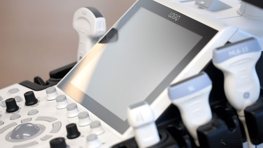
At Alpine BioMedical Clinic, we use ultrasound diagnostics as a supportive diagnostic tool to ensure ideal treatment planning and therapy. The LogiQ P10 by GE HealthCare is the device we use, which provides an outstanding representation of the internal organs and structures.

Med. pract. Dana Hreus M.A.

Frequently Asked Questions about Sonography and Ultrasound

In the following section, we will answer your questions in detail to provide you with more insight about sonography.

Sonography is an imaging procedure that uses high-frequency sound waves to create images of the body’s interior. These waves are known as ultrasound waves. This method is used to visualize organs, tissues, and blood vessels in detail and diagnose diseases without using harmful radiation.

There are various types of ultrasound, including:

  • B-Mode Ultrasound: This is the standard method that produces 2D images of tissues and organs.
  • Doppler Ultrasound: Used to measure blood flow velocity and detect vascular diseases.
  • 3D/4D Ultrasound: Creates detailed three-dimensional images and is commonly used during pregnancy.
  • Contrast-Enhanced Ultrasound: Uses contrast agents to better visualize blood vessels and organs.

During an ultrasound examination, a gel is applied to the skin for better sound transmission. The doctor then moves a transducer (ultrasound probe) over the area being examined. The transducer sends out ultrasound waves that are reflected by the tissue. These reflected waves are converted into images that are displayed on a monitor.

Ultrasound works by sending high-frequency sound waves through the body. These waves are reflected differently by tissues depending on their density. The ultrasound transducer receives these reflections and converts them into digital images that are displayed on a monitor. This makes it possible to visualize the structure and condition of organs or tissues.

Various organs and body parts can be examined with ultrasound, including the liver, bladder, lungs, heart, thyroid, and head. The abdominal area (e.g., kidneys, stomach, or intestines) can also be closely examined using an abdominal ultrasound.

Ultrasound describes the technology that uses high-frequency sound waves to create images of internal body structures. Sonography is the medical term for the application of this technology to diagnose and examine organs and tissues. However, the terms are often used interchangeably.

For a sonography examination, certain things should be considered to ensure accurate results. Depending on the body area being examined, you may need to fast, particularly for abdominal or liver examinations. Before a bladder examination, drink plenty of water to ensure the bladder is well-filled. Wearing loose-fitting clothing can also facilitate access to the areas being examined.

Sonography is performed by various medical specialists, depending on the body part being examined. Generally, general practitioners, internists, urologists, or gynecologists perform sonographies. For specific examinations such as ultrasound of the abdomen or liver, gastroenterologists may also be consulted. Sometimes radiologists also perform sonographies.

Further information

Further information intended to give a better overview of the topic.