Dark Field Microscopy
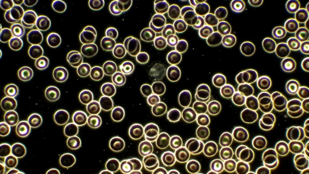
Dark field microscopy is a technique used to magnify and analyze the patient’s blood in a dark field microscope, therefore this method is also referred to as dark field analysis. For the examination, a small drop of blood is taken from the fingertip or earlobe, placed on a cover glass, and placed on the microscope slide. A thin blood film (a few micrometers thick) is formed between the slide and cover glass, in which the blood cells do not overlap and can be viewed individually.
Dark field microscopy has significant advantages over the classical blood smear, which represents dried and stained blood. In dark field analysis, the unaltered, living blood is displayed. The moving blood cells and plasma provide information about various aspects of the patient’s health condition.
Dr. med. Karsten Ostermann M.A.
At Alpine BioMedical Clinic, we use dark field microscopy as a supportive diagnostic tool. To achieve a sharp and precise visualization of the structures, high-quality optics are an absolute requirement for dark field microscopes.

Further information
Further information intended to give a better overview of the topic.